Understandings:
● Nucleosomes help to supercoil the DNA.
● DNA structure suggested a mechanism for DNA replication.
● DNA polymerases can only add nucleotides to the 3 end of a primer.
● DNA replication is continuous on the leading strand, and discontinuous on the lagging strand.
● DNA replication is carried out by a complex system of enzymes.
● Some regions of DNA do not code for proteins but have other important functions.
NATURE OF SCIENCE
Making careful observations: Rosalind Franklin’s X-ray diffraction provided crucial evidence that DNA is a double helix.
Applications and skills:
Guidance:
- DNA structure
To understand the detailed structure of DNA, you must be familiar with the numbering of the carbon atoms in the pentose sugar of DNA, which is deoxyribose.
- How is a single chain of DNA made up?
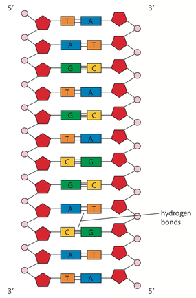
Each strand of DNA is composed of a backbone of alternating phosphate and deoxyribose molecules. These two molecules are held together by a covalent bond called a phosphodiester bond or linkage. A phosphodiester bond in DNA forms between a hydroxyl group of the 3ʹ carbon of deoxyribose and the phosphate group attached to the 5ʹ carbon of deoxyribose.
So, each nucleotide is attached to the previous one by this type of bond. This produces a chain of DNA. The reaction between the phosphate group on the 5ʹ carbon and the hydroxyl group on the 3ʹ carbon is a condensation reaction,
with a molecule of water released. When two nucleotides unite in this way, the two-unit polymer still has a 5ʹ carbon free at one end and a 3ʹ carbon free at the other end. Each time a nucleotide is added, it is attached to the 3ʹ carbon end. Even when thousands of nucleotides are involved, there is still a free 5ʹ carbon end with a phosphate group attached and a free 3ʹ carbon end with a hydroxyl group attached. This creates the alternating sugar–phosphate backbone of each chain.
As nucleotides are linked together with covalent phosphodiester bonds, a definite sequence of nitrogenous bases develops. This sequence carries the genetic code that is essential for the life of the organism.
- How are the two strands of DNA held together?
The two sugar–phosphate backbones are attached to each another by their nitrogenous bases. The two backbones
or chains run in opposite directions and are described as antiparallel. One strand has the 5ʹ carbon on the top and the 3ʹ carbon on the bottom; the other strand is the opposite way round
● Nucleosomes help to supercoil the DNA.
● DNA structure suggested a mechanism for DNA replication.
● DNA polymerases can only add nucleotides to the 3 end of a primer.
● DNA replication is continuous on the leading strand, and discontinuous on the lagging strand.
● DNA replication is carried out by a complex system of enzymes.
● Some regions of DNA do not code for proteins but have other important functions.
NATURE OF SCIENCE
Making careful observations: Rosalind Franklin’s X-ray diffraction provided crucial evidence that DNA is a double helix.
Applications and skills:
- Application: Rosalind Franklin and Maurice Wilkin’s investigation of DNA by X-ray diffraction.
- Application: Use of nucleotides containing dideoxyribonucleic acid to stop DNA replication in preparation of samples for base sequencing.
- Application: Tandem repeats are used in DNA profiling.
- Skill: Analysis of results of the Hershey and Chase experiment to provide evidence that DNA is the genetic material.
- Skill: Utilization of molecular visualization software to analyse the association between protein and
DNA within a nucleosome.
Guidance:
- Details of DNA replication differ between prokaryotes and eukaryotes. Only the prokaryotic system is expected.
- The protein enzymes involved in DNA replication should include helicase, DNA gyrase, single strand binding proteins, DNA primase, and DNA polymerases I and III.
- The regions of DNA that do not code for proteins should be limited to regulators of gene expression, introns, telomeres, and genes for tRNAs
- DNA structure
To understand the detailed structure of DNA, you must be familiar with the numbering of the carbon atoms in the pentose sugar of DNA, which is deoxyribose.
- How is a single chain of DNA made up?
Each strand of DNA is composed of a backbone of alternating phosphate and deoxyribose molecules. These two molecules are held together by a covalent bond called a phosphodiester bond or linkage. A phosphodiester bond in DNA forms between a hydroxyl group of the 3ʹ carbon of deoxyribose and the phosphate group attached to the 5ʹ carbon of deoxyribose.
So, each nucleotide is attached to the previous one by this type of bond. This produces a chain of DNA. The reaction between the phosphate group on the 5ʹ carbon and the hydroxyl group on the 3ʹ carbon is a condensation reaction,
with a molecule of water released. When two nucleotides unite in this way, the two-unit polymer still has a 5ʹ carbon free at one end and a 3ʹ carbon free at the other end. Each time a nucleotide is added, it is attached to the 3ʹ carbon end. Even when thousands of nucleotides are involved, there is still a free 5ʹ carbon end with a phosphate group attached and a free 3ʹ carbon end with a hydroxyl group attached. This creates the alternating sugar–phosphate backbone of each chain.
As nucleotides are linked together with covalent phosphodiester bonds, a definite sequence of nitrogenous bases develops. This sequence carries the genetic code that is essential for the life of the organism.
- How are the two strands of DNA held together?
The two sugar–phosphate backbones are attached to each another by their nitrogenous bases. The two backbones
or chains run in opposite directions and are described as antiparallel. One strand has the 5ʹ carbon on the top and the 3ʹ carbon on the bottom; the other strand is the opposite way round
- DNA packaging
The DNA molecules of eukaryotic cells are paired with a type of protein called histone. Actually, there are several histones, and each helps in DNA packaging. Packaging is essential because the nucleus is microscopic and a single human molecule of DNA in a chromosome may be 4 cm long.
When looking at unfolded DNA with an electron microscope, you can see what looks like beads on a string. Each of the beads is a nucleosome. A nucleosome consists of two molecules of each of four different histones. The DNA wraps twice around these eight protein molecules. The DNA is attracted to the histones because DNA is negatively charged and histones are positively charged. Between the nucleosomes is a single string of DNA. There is often a fifth type of histone attached to the linking string of DNA near each nucleosome. This fifth histone leads to further wrapping (packaging) of the DNA molecule, and eventually to highly condensed or supercoiled chromosomes.
When DNA is wrapped around the histones and then further wrapped in even more elaborate structures, it is inaccessible to transcription enzymes. Therefore, the wrapping or packaging of DNA brings about a regulation of the transcription process. This allows only certain areas of the DNA molecule to be involved in protein synthesis.
- Types of DNA sequences
The genomes, complete DNA sequences, of many eukaryotes are now known because of rapid advancements in the field of genomics. Genomics involves the science of sequencing, interpreting, and comparing whole genomes. From the multinational Human Genome Project, we have learned that less than 2% of human DNA actually codes for proteins or other materials required for protein synthesis in the cell.
Regions of DNA that do not code for proteins include areas that act as regulators of gene expression, introns, telomeres, and genes that code for transfer ribonucleic
acids (tRNAs).
- Structural DNA
Structural DNA is highly coiled DNA that does not have a coding function. It occurs around the centromere and near the ends of chromosomes at the telomeres. Some scientists refer to the sections of DNA that appear to not have a coding function as pseudogenes. Many feel these sections have lost their function due to a mutation involving a base sequence change.
- DNA replication
When Watson and Crick proposed their model for the structure of DNA, they realized that the A–T and C–G base pairing provided a way for DNA to be copied. They thought that a single strand of DNA could serve as a template for a copy. This would facilitate the accuracy that is necessary to pass on the DNA information from one generation to the next. They referred to this idea as a semi-conservative model of DNA replication. The experiments carried out in the late 1950s by Matthew Meselson and Franklin Stahl confirmed this DNA semi-conservative model of replication.
- Semi-conservative replication
The process of DNA replication involving bacteria was developed as a result of Meselson and Stahl’s experiment. The replication of DNA begins at special sites called origins of replication. Bacterial DNA is circular, has no histones, and has a single origin. Eukaryotic DNA is linear, has histones, and has thousands of origins. The presence of multiple replication origins greatly accelerates the copying of large eukaryotic chromosomes.
Here is a brief summary of the replication process
- Replication begins at the origin, which appears as a bubble because of the separation of the two strands. The separation or ‘unzipping’ occurs because of the action of the enzyme helicase on the hydrogen bonds between nucleotides.
- At each end of a bubble there is a replication fork. This is where the double- stranded DNA opens to provide the two parental DNA strands that are the templates necessary to produce the daughter DNA molecules by semi- conservative replication.
- The bubbles enlarge in both directions, showing that the replication process is bidirectional. The bubbles eventually fuse with one another to produce two identical daughter DNA molecules.

- Elongation of a new DNA strand
The production of a new strand of DNA using the templates exposed at the replication forks occurs in an orderly manner.
- Replication proteins
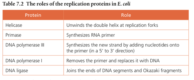
The basic processes of DNA replication were worked out with research using E. coli. Table 7.2 summarizes the roles of the replication proteins in E. coli.
The production of a new strand of DNA using the templates exposed at the replication forks occurs in an orderly manner.
- A primer is produced under the direction of primase at the replication fork. This primer is a short sequence of RNA, usually only 5–10 nucleotides long. Primase allows the joining of RNA nucleotides that match the exposed DNA bases at the point of replication.
- The enzyme DNA polymerase III then allows the addition of nucleotides in a 5ʹ to 3ʹ direction to produce the growing DNA strand.
- DNA polymerase I also participates in the process. It removes the primer from the 5ʹ end and replaces it with DNA nucleotides.
- Replication proteins
The basic processes of DNA replication were worked out with research using E. coli. Table 7.2 summarizes the roles of the replication proteins in E. coli.